Gang Chen1-3 ![]() ,
Min Feng2,3
,
Min Feng2,3
For correspondence:- Gang Chen Email: gangch_tcm@163.com Tel:+862362769785
Received: 12 March 2015 Accepted: 9 April 2016 Published: 28 June 2016
Citation: Chen G, Feng M. Ameliorative effect and potential mechanism of Ermiao san on adjuvant-induced arthritis in rats. Trop J Pharm Res 2016; 15(6):1159-1165 doi: 10.4314/tjpr.v15i6.7
© 2016 The authors.
This is an Open Access article that uses a funding model which does not charge readers or their institutions for access and distributed under the terms of the Creative Commons Attribution License (http://creativecommons.org/licenses/by/4.0) and the Budapest Open Access Initiative (http://www.budapestopenaccessinitiative.org/read), which permit unrestricted use, distribution, and reproduction in any medium, provided the original work is properly credited..
Purpose: To investigate the effect and mechanism of action of Ermiao san (EMS), a traditional Chinese herbal formula, on inflammation development and production of inflammatory mediators in adjuvant-induced arthritis (AIA).
Methods: AIA was induced by injection of 0.1 ml Freund’s complete adjuvant (FCA, 10 mg/ml) in the left hind footpad of the rats. AIA rats were intragastricly treated with 0.5, 1, 2 g/kg EMS or 0.1 g/kg methotrexate from day 7 to 28 after FCA challenge. Foot volume and histological score were measured. Osteoclast number was calculated by tartrate-resistant acid phosphatase (TRAP) staining assay. Levels of prostaglandin (PG) E2, tumor necrosis factor (TNF) -α and interleukin (IL)-1β in serum were determined by enzyme-linked immunosorbent assay (ELISA) while the level of nitric oxide (NO) in serum was analyzed by Griess reaction method.
Results: Foot volume, histological score, osteoclast number and serum levels of TNF-α, IL-1β, PGE2 and NO were all increased in AIA group rats on day 28 after FCA challenge (all p < 0.01) compared with control. EMS (1 and 2 g/kg) significantly decreased the foot volume of AIA rats by 10 % (p < 0.05) and 19 % (p < 0.01), respectively, compared with AIA group. Furthermore, 1 and 2 g/kg EMS significantly reduced histological score by about 28 % (p < 0.05) and 46 % (p < 0.01), respectively, as well as osteoclast number by 12 % (p < 0.05) and 15 % (p < 0.05), respectively, compared with AIA group. In addition, 1 and 2 g/kg EMS significantly decreased the serum levels of TNF-α about 23 % (p < 0.05) and 43 % (p < 0.01), IL-1β by15 % (p < 0.05) and 26 % (p < 0.01), NO 13 % (p < 0.05) and 26 % (p < 0.01) as well as PGE2 by 11 % (p < 0.05) and 15 % (p < 0.01), respectively, compared with AIA group.
Conclusion: These results suggest that EMS probably alleviates arthritis development and joint destruction by decreasing the production of inflammatory mediators in AIA rats.
Introduction
Rheumatoid arthritis (RA) is an autoimmune disease characterized by chronic inflammation with the lesions of cartilage and bone, and finally results in joint destruction and deformity [1]. Inflammatory mediators such as interleukin (IL)-1β, tumor necrosis factor (TNF)-α, prostaglandin E2 (PGE2) and nitric oxide (NO) play a key role in the pathogenesis of RA [2-4]. Antigen-presenting cells, including dendritic cells, macrophages and B cells, present RA-associated antigens to T cells [5]. Simultaneously, antigen-activated CD4+ T cells amplify the immune response by activating multiple types of cells including synovial fibroblasts, T cells, B cells, monocytes, macrophages, chondrocytes and osteoclasts [6]. These activated cells can produce a variety of inflammatory mediators which contribute to the synovitis development. Concurrently, inflammatory mediators may stimulate inflammatory cells to produce more inflammatory mediators, as well as matrix metalloproteinases, aggrecanases and collagenases which contribute to the destruction of cartilage and bone [7].
In current clinical practice, non-steroidal anti-inflammatory drugs, corticosteroids, disease-modifying anti-rheumatic drugs and biologic agents are the main anti-rheumatic drugs applied for RA treatment [8]. However, severe adverse and toxic effects, expensive costs and limited efficacy spur RA patients on to natural herbal therapies including traditional Chinese medicine (TCM) for its effectiveness and hypotoxicity in recent years [9].
Ermiao san (EMS) is a traditional Chinese herbal formula which is composed of Atractylodes chinensis (DC.) Koidz. and Phellodendron chinensis Schneid. (Rutaceae). in a weight ratio of 1:1. EMS has been empirically used to treat “Bi Zheng” for hundreds of years, and in current clinic of TCM, EMS and its modified formulae are frequently used for treatment of RA [10]. Documents have demonstrated the pharmacological activities of EMS. EMS showed a remarkable improvement against the elevation in serum transaminase levels as well as in histopathological changes of immunological liver injury in mice [11]. Chinese herbal formulas Si-Wu-Tang and EMS synergistically ameliorated hyperuricemia and renal impairment in rats [12]. However, up till now, the effect and potential mechanisms of EMS on adjuvant-induced arthritis (AIA) in rats are not totally understood. Therefore, in the present study, we investigated the effect of EMS on the arthritis development and production of inflammatory mediators in AIA of rats.
Methods
Preparation of EMS extract
Atractylodes chinensis (DC.) Koidz. and Phellodendron chinensis Schneid. (Rutaceae) were purchased from Chongqing Tongjunge Pharmacy (Chongqing, China) and were identified by Dr. Jifen Zhang, College of Pharmaceutical Sciences, Southwest University (Chongqing, China). Morphological, microscopic authentications and thin layer chromatography were performed in accordance to Chinese Pharmacopoeia (2010). Herbarium voucher specimens of the tested herbs were deposited at the Chongqing Key Laboratory of Nature Medicine Research, Chongqing Technology and Business University, with voucher specimen numbers as follows: 2013-109 (Atractylodes chinensis (DC.) Koidz.) and 2013-187 (Phellodendron chinensis Schneid. (Rutaceae)). Raw herbal materials (500 g:500 g) were extracted with boiling water (1:8, w / v) for 1 h and the extraction was repeated twice. The solution was filtered and concentrated and then made into freeze-dried powder. 268 g EMS freeze-dried extract powder was obtained from 1000 g raw material (the yield was 26.8 %). The freeze-dried powder was stored at -70 °C until use.
Drugs and chemicals
All chemicals and reagents were purchased from Sigma-Aldrich (USA) unless otherwise specified. Enzyme-linked immunosorbent assay (ELISA) kits for TNF-α, IL-1β and PGE2 were purchased from Pierce (USA). Bacillus Calmette-Guerin (BCG) was purchased from Shanghai biochemical factory (China). Methotrexate (MTX) was purchased from Shanghai Xinyi pharmaceutical factory (China). Tartrate-resistant acid phosphatase (TRAP) kit was purchased from Sigma.
Induction of AIA
Male Sprague-Dawley rats weighing 200-220 g were purchased from the Third Military Medical University (China). All rats were housed in a temperature-controlled room (22 ± 2 °C) under a light/dark cycle with lights on from 7:00 am to 7:00 pm. They were allowed food and water ad libitum. All animal procedures were approved by the Ethical Committee in Animal Research of Chongqing Technology and Business University (ref no. 2013-7-12/CTBU). Freund’s complete adjuvant (FCA) was prepared by suspending heat-killed BCG in sterile mineral oil (10 mg/ml). Arthritis was induced by a single injection of 100 μl of FCA intradermally in the left hind footpad of each rat [13].
Drug therapy
Rats were randomly divided into six groups with 8 rats in each group: (1) Control group, (2) AIA group, (3) 0.1 mg/kg MTX group, (4) 0.5 g/kg EMS group, (5) 1 g/kg EMS group, (6) 2 g/kg EMS group. According to the clinical practice of TCM, the dosage of EMS for adults (60 kg/person) is 30 g/60 kg/d, equivalently, for rats, this dosage is 3 g/kg/d calculated by the formula that converts dosage of human into that of rats according to the respective body surface areas.
In the present study, the yield of EMS water extract was 26.8 %, making the dosage of XTW extract about 1 g/kg/d. Arthritic rats of (3), (4), (5) and (6) groups were intragastricly treated with MTX or EMS once a day. For the Control and AIA groups, rats were intragastricly treated with 2 ml physiological saline. All the rats received treatment from day 7 to day 28 after FCA immunization.
Assessment of foot volume
From day 7 to day 28 following FCA challenge, foot volume of the left hind of all the rats was measured by PBC7140 plethysmometer (Ugo Basile, Italy) every 7 days [14]. The measurement was carried on by one investigator blind to the experimental protocol.
Assessment of histological score
All rats were sacrificed 1 h after the last drug treatment. The left ankle joints were surgically removed, fixed in 4 % paraformaldehyde for 48 h, decalcified in 10 % ethylene diamine tetraacetic acid for 28 days, then embedded in paraffin. Serial paraffin sections (6 μm) were stained with hematoxylin and eosin (H&E). Histopathological changes in joint were scored (histological score) using the following parameters by one investigator blind to the experimental protocol [15]: 0: normal, 1: infiltration of inflammatory cells, 2: synovial hyperplasia, 3: pannus formation, 4: bone erosion, 5: bone destruction.
Assessment of osteoclast number
Serial paraffin sections were prepared as previously described in the section of “Assessments of histological score”. TRAP staining was carried on according to the manufacturer’s instructions. Briefly, tissue sections were dewaxed in xylene and rehydrated in graded ethanol. After washing with distilled water, sections were incubated in the reaction mixture at 37 °C in a humid and light-protected incubator for 1 h. Thereafter, sections were washed three times with distilled water, counterstained with hematoxylin, and observed under a microscope. The number of osteoclasts (TRAP-positive cells) was calculated at high magnification (× 100). The results are expressed as the total number of osteoclasts per slide [16].
Analysis of inflammatory mediators
Blood was obtained by cardiac puncture under ether anesthesia prior to sacrifice. Serum was obtained from coagulated blood by centrifuge at 4000 g for 20 min. Serum samples were stored at -20 °C until use. Levels of TNF-α, IL-1β and PGE2 in serum were tested using enzyme-linked immunosorbent kits. Level of NO in serum was determined with a NO test kit based on the Griess reaction method. Assays were performed according to the manufacturer’s instructions.
Statistical analysis
All data were presented as means ± standard error (SD). Statistical comparisons were evaluated by ANOVA test using SPSS 16 software. Differences between values were considered significant at p < 0.05.
Results
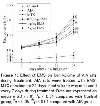
Effect of EMS on foot volume of AIA rats
Arthritis had developed in the immunized rats on the 3rd day after FCA challenge. Joint swelling and erythema were observed as obvious signs of the evolution of arthritis. There was no significant difference in the arthritis amongst all the groups (two groups of AIA, MTX and three doses of EMS groups) on day 7. However, following treatment, compared with the control group, foot volume of rats in AIA group was markedly enlarged on day 28 (p < 0.01, ). On treatment with 1 and 2 g/kg EMS for 21 days, foot volume of arthritic rats were significantly decreased compared with AIA group given saline (p < 0.05 and p < 0.01 respectively, ). However, treatment with 0.5 g/kg EMS for 21 days did not statistically reduce the foot volume (). Treatment with 0.1 mg/kg MTX for 21 days remarkably decreased the foot volume compared with AIA group (p < 0.01, ).
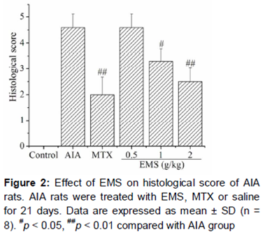
Effect of EMS on histological score of AIA rats
Histological score represents the degree of severity of articular inflammation and destruction. Compared with Control group, histological score of AIA group was markedly increased on day 28 (p < 0.01, ). Treatment with 1 and 2 g/kg EMS significantly reduced the histological score compared with AIA group in a dose-dependent manner (p < 0.05 and p < 0.01 respectively, ). 0.1 mg/kg MTX also markedly decreased the histological score (p < 0.01, ). However, 0.5 g/kg EMS did not statistically reduced the histological score compared with AIA group.
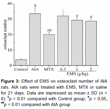
Effect of EMS on osteoclast number of AIA rats
To evaluate the effect of EMS on osteoclast number, joint sections were stained with TRAP. A significant increase of osteoclast number was observed in AIA group compared with the Control group (p < 0.01, ). Treatment with 1 and 2 g/kg EMS significantly reduced the osteoclast number compared with AIA group (both p < 0.05, ). Meanwhile, 0.1 mg/kg MTX also markedly decreased the osteoclast number compared with AIA group (p < 0.01, ). However, a statistical reduction of osteoclast number was not observed in 0.5 g/kg EMS group compared with AIA group.
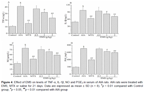
Effects of EMS on levels of inflammatory mediators in serum of AIA rats
The levels of TNF-α, IL-1β, PGE2 and NO in serum were all significantly elevated in AIA group rats than in Control group rats (p < 0.01, ). Treatment with 1 and 2 g/kg EMS markedly decreased the serum levels of TNF-α, IL-1β, PGE2 and NO compared with Control group in a dose-dependent manner (p < 0.05 and p < 0.01 respectively, ). Meanwhile, 0.1 mg/kg MTX significantly decreased the serum levels of TNF-α, IL-1β, PGE2 and NO compared with AIA group rats (p < 0.01, ).However, 0.5 g/kg EMS did not statistically reduced the serum levels of TNF-α, IL-1β, PGE2 and NO compared with AIA group rats.
Discussion
In this study, we demonstrated that EMS, a traditional Chinese herbal formula, significantly decreased the foot volume, histological score and osteoclast number in arthritic tissues of adjuvant-induced arthritis (AIA) rats. Simultaneously, treatment with EMS significantly reduced the levels of TNF-α, IL-1β, PGE2 and NO in serum of AIA rats.
It is well-known fact that redness, swelling, heat and pain are the main characteristics of inflammation [13]. In AIA, a classic animal model of human rheumatoid arthritis (RA), foot volume is verified to be closely related to arthritis development. Currently, foot volume is one of the most frequent-used indices to measure the anti-arthritic effect of tested drugs [17].
In this study, foot volume was markedly increased after intradermal injection of FCA for 28 days in rats. After treatment with EMS for 21 days, foot volume of AIA rats were significantly reduced in a dose-dependent manner. At the same time, we also found that both erythralgia and dysfunction in EMS-treated AIA rats were clearly alleviated. These results suggested that EMS might significantly attenuate the inflammatory development of AIA in rats.
It is reported that RA is the leading cause of disability in the world due to the progressive erosion of cartilage and bone. Numerous evidences suggest that joint destruction in RA is mainly due to chronic inflammation [18]. In RA, the synovial tissue undergoes hypertrophy via proliferation of the synovial cells and homing of monocytes and fibroblasts, and the result is development of the rheumatoid pannus [19]. Rheumatoid pannus is considered as the hallmark of chronic inflammation and mediates cartilage damage in RA. Many inflammatory mediators such as IL-1β and TNF-α, can activate the osteoclasts [20]. The abundant activated osteoclasts that are characteristic of rheumatoid inflammation directly destroy bone [21]. Histological score and osteoclast number are two frequently-used index which represent the degree of severity of articular inflammation and destruction. In this study, the histological score and osteoclast number were significantly increased in AIA rats which indicated the serious joint destruction induced by FCA in the rats. However, treatment with EMS dose-dependently reduced the histological score and osteoclast number, which suggested that EMS might significantly alleviate the joint destruction in AIA rats.
A great number of clinical and experimental data support the pivotal role of TNF-α, IL-1β, PGE2 and NO in the pathogenesis of RA. TNF-α promotes bone resorption directly through activation of cells of the osteoclasts [22], and indirectly through the expression of osteoclast activators [23]. TNF-α also suppresses bone formation via increased osteoblast apoptosis [24], and reduced differentiation and proliferation of osteoblasts and their progenitors [25]. IL-1β reduces chondrocyte proteoglycan synthesis [26], increases the synthesis of matrix metalloproteinases and the release of nitric oxide [27]. Transgenic mice deficient in IL-1 receptor antagonist spontaneously develop a chronic arthritis similar to RA with bone erosions and present higher susceptibility for collagen-induced arthritis [28]. Other studies show that IL-1β is not predominant in the acute inflammatory stages of most experimental arthritis models, but plays a significant role in perpetuating joint inflammation and in the pathogenesis of bone and cartilage damage [29]. NO contributes to T cell activation in RA by altering multiple signaling pathways in T cells [30]. NO produced in the inflammatory joint may contribute to the peri-articular bone-loss observed in RA. Transgenic mice deficient in NO production are resistant to IL-1-induced bone resorption [31]. PGE2 is considered to be the major contributor to inflammatory pain in RA. PGE2 and NO also promote inflammation development and participate in destructive mechanisms in the rheumatoid joint [32]. These evidences highlight the key role of TNF-α, IL-1β, PGE2 and NO in the inflammation development and bone and cartilage damage in RA. In this study, levels of TNF-α, IL-1β, PGE2 and NO in serum of AIA rats were all significantly elevated, which suggested the important role of these inflammatory mediators in the pathogenesis of AIA in rats. Treatment with EMS significantly lowered the levels of TNF-α, IL-1β, PGE2 and NO in serum of AIA rats, although the degree of reduction were lower than methotrexate, the positive controlled drug used in this study. These findings suggest that the ameliorative effect of EMS on foot volume and joint destruction may be closely related to the reduction of production of TNF-α, IL-1β, PGE2 and NO in AIA rats.
Conclusion
The findings of the study indicate that EMS suppresses inflammation development and joint destruction in AIA rats, and that the potential mechanism may be closely associated with inhibition of the production of inflammatory mediators including TNF-α, IL-1β, PGE2 and NO. These data provide mechanistic evidence for anti-arthritic application of EMS in TCM and also suggest that EMS is a promising candidate for novel therapeutic agents for RA.
Declarations
Acknowledgement
References
Archives


News Updates